
External ear - made up of 5 parts:
1. auricle (aka "pinna"):
 |
| The auricle or pinna refers tot he entire visible structure of the External ear (i.e. does not include the External acoustic meatus). |

3. lobule:

4. external acoustic meatus (aka "auditory canal"):
 |
| Anterior view of External acoustic meatus. |
 |
| Lateral view of External acoustic meatus. |
5. tympanic membrane:

Middle ear - made up of 4 parts:


A. Oval window:
 |
| Oval window is the hollow circle formed by the "footplate" or base of the Stapes, which is broken on this model. |
B. Round window:

C. Auditory tube (aka "Pharyngotympanic tube"):

D. Auditory ossicles (3 "little bones"):
i. malleus:

ii. incus:
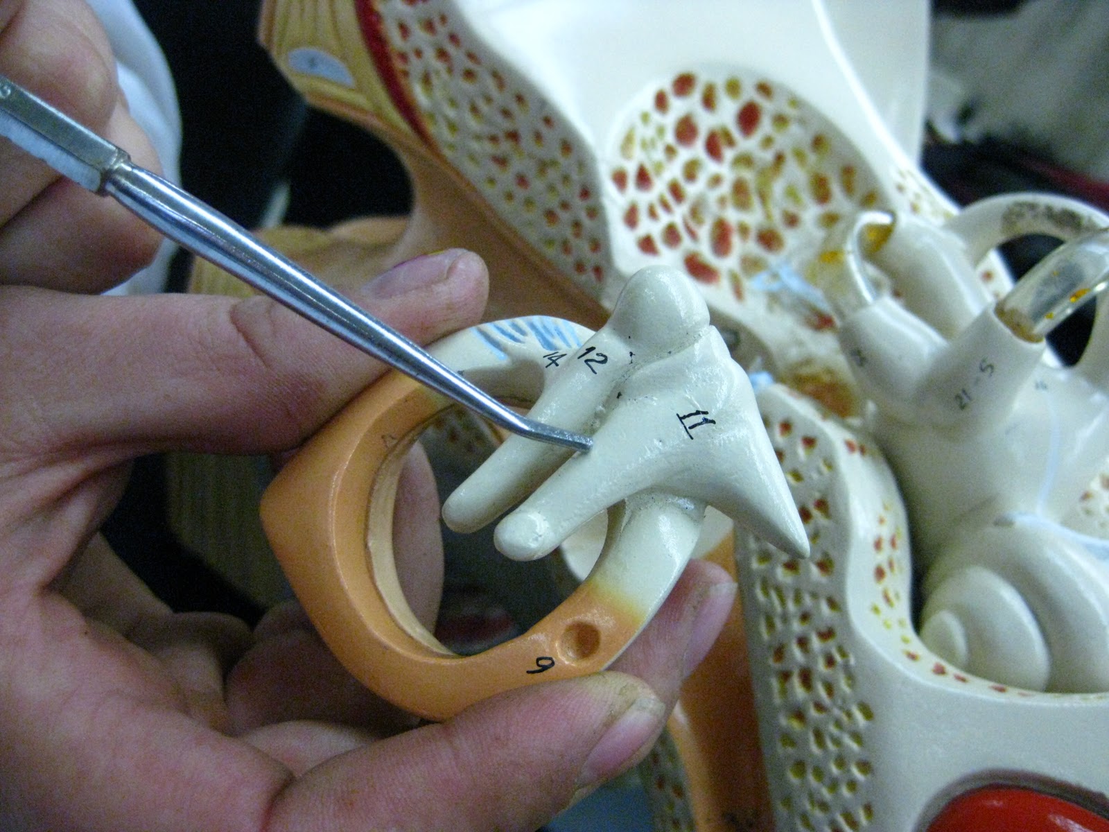
iii. stapes: no picture available (stapes broken on model). Link to clear picture of stapes on a different ear model.
Internal ear (aka "Inner ear" or "Labyrinth") - made up of 2 separate labyrinths:
1. Osseous labyrinth (aka "Bony labyrinth"):
 |
| Bony labyrinth contains the fluid called "perilymph". |
2. Membranous labyrinth:
 |
| Membranous labyrinth contains the fluid called "endolymph". |
1. vestibule (of Bony Labyrinth) - contains 2 internal structures of interest.
 |
| The Vestibule of the inner ear connects the Cochlea with the Semicircular canals. |
i. utricle (of vestibule, of Membranous labyrinth): one of two small cavities inside the vestibule; not visible on the model because it is an internal structure. Internet diagram of the utricle can be found here.
ii. saccule (of vestibule, of Membranous labyrinth): one of two small cavities inside the vestibule. See "utricle" entry above for a diagram of the saccule.
2. Semicircular Canals (of Bony Labyrinth, 3 total) - contains 1 internal structure of interest.
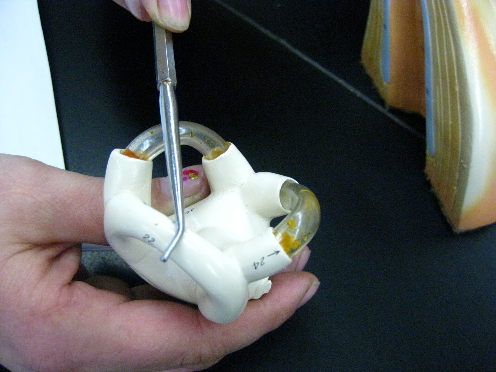
 |
| Ampullae are dilated (enlarged) areas of the base of each Semicircular canal. |
3. Cochlea - (of Bony labyrinth, made up of 3 internal tubes):
 |
| Cochlea is a "snail-like" structure dedicated to the sense of hearing (whereas the Vestibule and Semicircular canals are dedicated to balance/spatial orientation). |
i. scala vestibuli (of Cochlea, of Bony labyrinth):
 |
| Scala vestibuli are Orange on this model. Scala vestibuli is filled with "perilymph". |
ii. scala tympani (of Cochlea, of Bony labyrinth):
 |
| Scala tympani are Blue on this model. Scala tympani is filled with "perilymph". |
iii. scala media (of Cochlea, of Membranous labyrinth, aka "Cochlear Duct"):
 |
| Scala media (aka "Cochlear Duct") is filled with "endolymph". |
Vestibulocochlear Nerve (aka "Cranial Nerve 8"):

No comments:
Post a Comment